
Early detection is the most important factor in successfully treating skin cancer. At Koncept Medical Clinic, we offer professional Skin Cancer Screening and Mole Assessment to help identify suspicious lesions early, provide medical guidance and ensure timely referral where appropriate. Our clinicians are trained in assessing moles, freckles, sun spots and other skin lesions, offering reassurance and safe clinical evaluation in a CQC-registered medical environment.
Whether you have noticed a new mole, a change in an existing lesion, or simply want a routine skin check, we provide thorough and confidential assessments for adults of all ages.
Skin Cancer Screening involves a detailed examination of the skin to identify signs of:
Melanoma
During screening, we assess:
Size, shape and colour of moles
While many lesions turn out to be harmless, early detection of skin cancer is crucial.
Patients choose medical skin checks because they offer:
Early diagnosis significantly improves outcomes.
Your clinician examines moles using recognised dermatology guidelines (e.g., ABCDE).
Most lesions are harmless — screening offers clarity.
Helpful for long-term skin protection.
Reducing uncertainty and anxiety.
Knowing what changes to watch for between appointments.
Including NHS referral or private dermatology pathways.
Skin cancer is one of the most treatable forms of cancer when detected early.
You should book a screening if you notice:
A mole changing in size, colour or shape
A new mole appearing in adulthood
A mole with irregular, blurred or jagged borders
A lesion that bleeds, crusts or doesn’t heal
A patch of skin that is persistently scaly
A mole that becomes itchy or painful
A flat red or rough patch that keeps returning
A firm, pearly or ulcerated lesion
High-risk groups include:
Individuals with fair skin or freckles
People with many moles
Family history of melanoma
Previous sunburn or tanning bed exposure
Outdoor workers
History of skin cancer
Urgent assessment recommended if:
A mole changes rapidly
There is obvious asymmetry or irregularity
A lesion bleeds without cause
A sore does not heal after 4–6 weeks
If a lesion looks suspicious, we will advise immediate referral for specialist assessment.
These symptoms require immediate medical attention. Do not wait for a clinic appointment.
Your clinician will discuss:
Your concerns
Personal and family history
Sun exposure habits
Any previous skin cancers
Changes you’ve noticed in specific lesions
We then proceed with a careful skin examination.
Your clinician examines:
All areas of concern
Sun-exposed areas (face, neck, arms, chest)
Any moles you point out for review
Additional areas if clinically relevant
We use dermatology assessment criteria such as:
ABCDE Method for Moles
Asymmetry
Border irregularity
Colour variation
Diameter
Evolving changes
We also check for features of basal cell or squamous cell carcinoma.
Following examination, you will receive clear guidance:
If the lesion appears benign:
Reassurance
Advice on self-monitoring
Sun safety recommendations
Optional routine follow-up
If the lesion requires further assessment:
We will recommend referral to:
Your NHS GP for urgent dermatology pathway
or
Private dermatology (if you prefer)
We ensure you understand next steps clearly and safely.
You may be advised to:
Take regular photographs of moles
Monitor changes
Wear high-factor SPF daily
Avoid excessive sun exposure
Schedule routine annual skin checks (high-risk patients only)
We provide long-term reassurance and guidance.
• Profhilo: noticeable at 2–4 weeks, full effect at 8 weeks
• Polynucleotides: visible improvements from 3–6 weeks, progressive stimulation over time
A course of 2–3 sessions is usually recommended for optimal results.
CQC-registered medical clinic
Trained clinicians experienced in mole and lesion assessment
Safe, evidence-based clinical examination
Thorough, clear and supportive assessments
Fast access to onward referral if needed
Confidential and patient-centred care
Convenient Kingston location
Option to review additional skin concerns during the same appointment
KONCEPT® Medical Clinic proudly serves patients from across Kingston upon Thames and the surrounding areas, including:
Our clinic is conveniently located with easy transport links, making it simple to attend appointments and receive timely care when you need it.

Call To book
email to book


Targeted treatments to soothe inflammation, reduce flare-ups, and support long-term skin barrier health.

Specialist-led assessment and treatment for a wide range of skin concerns.
Look for changes in size, colour, shape or texture — or new symptoms like itching or bleeding.
Suspicious lesions require specialist referral for biopsy. We do not remove lesions that may be cancerous in a cosmetic setting.
High-risk patients (light skin, many moles, family history) benefit from yearly reviews.
Some sun damage can be improved with skincare and treatments; we will guide you safely.
We will advise you to see your NHS GP or a private dermatologist urgently for biopsy assessment.
No — it is a visual and clinical examination only.
Yes — these areas can also be examined if accessible.
Skin Cancer Screening at Koncept Medical Clinic provides professional, thorough assessment of moles, lesions and sun-exposed skin. With early detection and clear guidance, our clinicians offer reassurance, safety and support, helping you protect your skin health with confidence.
Skin cancer results from the abnormal growth of skin cells, often linked to UV radiation. At KONCEPT®, our specialists diagnose and treat the three main types of skin cancer and common pre-cancerous lesions:
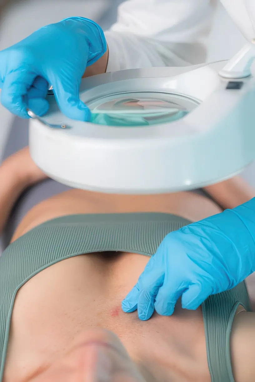
At KONCEPT Medical Clinic, we offer precise, meticulous screening exams, from full-body checks to focused evaluations, to precisely target your unique risk profile.
Basal Cell Carcinoma (BCC): Characterised by a pearly, waxy bump or a flat, flesh-colored or brown scar-like lesion; rarely spreads.
Squamous Cell Carcinoma (SCC): Characterised by a firm, red nodule or a flat, crusty lesion; can spread if left untreated.
Melanoma: The most serious form, often characterized by the ABCDE signs (Asymmetry, Border irregularity, Color variation, Diameter >6mm, Evolving); spreads aggressively.
Actinic Keratosis (AK): A pre-cancerous, scaly patch found on sun-exposed areas; the precursor to SCC.
There is no permanent cure for Rosacea, but with expert management, you can achieve long-term remission and significant symptom reduction. Our strategy integrates three key areas:
Your screening begins with a detailed medical consultation to understand your sun exposure history and identify specific risk factors (e.g., genetics, past severe sunburns, tanning bed use).
A meticulous examination by our dermatologist to detect subtle, concerning lesions often missed by self-examination.
(Asymmetry, Border irregularity, Color variation, Diameter, Evolving) is used to professionally assess all existing moles (nevi) for signs of Melanoma.
We determine your ideal follow-up frequency (e.g., every 6-12 months) based on your individual risk profile (e.g., number of moles, family history).
For ongoing skin health, we reinforce best practices for daily sun defense and prevention of new damage.
For confirmed or highly suspicious lesions, we rely on state-of-the-art technology to ensure precision, minimizing unnecessary procedures and providing a definitive diagnosis.
Utilized for highly focused, magnified evaluation of suspicious lesions, identifying subtle patterns invisible to the naked eye.
A complete photographic baseline map of your skin to help detect any new or changing moles during future follow-up exams.
A necessary, minor procedure to remove a small tissue sample for laboratory analysis (histopathology) to confirm or rule out cancer.
For confirmed cancerous lesions, we plan the surgical procedure to remove the skin cancer completely with clear margins, ensuring the best outcome.
While visual screening identifies potential concerns, advanced technology is the most effective way to accurately diagnose a lesion and ensure its complete, definitive removal.
We offer a spectrum of effective, evidence-based treatments and diagnostics, from minimally invasive biopsies to advanced surgical therapy:
Diagnosis (BCC, SCC, Melanoma)
A necessary, minor surgical procedure to remove a tissue sample for laboratory analysis (histopathology) to confirm the presence of cancer.
Treatment (BCC, SCC, Melanoma)
The standard, definitive treatment used to fully remove the cancerous lesion along with a margin of healthy surrounding tissue (clear margins).
Complex Cases (Face, Recurrence)
A specialized technique where the cancer is removed layer by layer and immediately checked under a microscope, preserving the maximum amount of healthy skin.
Treatment (Actinic Keratosis, Superficial BCC)
For pre-cancerous lesions or very superficial skin cancers, freezing (cryotherapy) or specialized creams can be used to destroy the abnormal cells.
Skin cancer can be serious, but it is manageable and highly curable when managed by experts who understand its subtle signs and complexity.
We offer the clinical resources and deep understanding required to correctly diagnose your lesion type (BCC, SCC, Melanoma) and tailor a risk-stratified surveillance plan.
We utilise industry-leading diagnostic tools and advanced surgical techniques (e.g., Digital Dermoscopy, Mohs Surgery) specifically calibrated for effective diagnosis and precise removal.
Skin cancer risk often requires lifelong vigilance. We are committed to a long-term partnership with you, setting optimal screening schedules and ensuring sustained prevention and skin health.
Call To book
email to book
Have a question? Chat with us on WhatsApp!