
At Koncept Medical Clinic, we provide safe, clinically led skin biopsy and histology services for lesions that require medical assessment. A biopsy involves removing a small sample of skin so it can be examined under a microscope by a specialist pathologist. This is the most accurate way to diagnose many skin conditions, including inflammatory skin disease and potential skin cancers.
Our clinicians will assess whether a biopsy is necessary, discuss the procedure with you in detail, and ensure the sample is processed through an accredited laboratory for a reliable diagnosis. We offer discreet, supportive care at every step, helping you understand your results and next steps clearly.
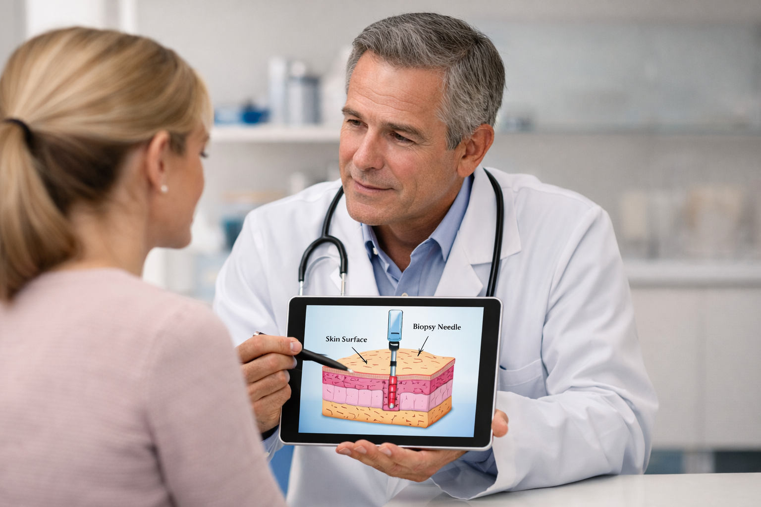
A skin biopsy is a minor medical procedure performed under local anaesthetic to remove a small sample of skin for laboratory testing. It is used to diagnose:
Suspicious moles
The sample is sent to a histology laboratory, where a specialist examines the tissue under a microscope to identify any abnormal cells or confirm a diagnosis.
Biopsies are essential when visual assessment alone isn’t enough to determine the nature of a lesion.
Patients choose biopsy assessment because it provides:
Clinical examination alone cannot always confirm the cause — histology provides clarity.
If skin cancer is suspected, early diagnosis is crucial.
Many lesions turn out to be benign; biopsy provides reassurance.
Histology results direct the safest and most effective next steps.
Samples are examined by qualified dermatopathologists.
Helps reduce anxiety about changing or unusual lesions.
A biopsy is the gold standard for diagnosing many skin conditions.
You may need a biopsy if you have:
A mole that is changing in size, shape, or colour
A new lesion appearing in adulthood
A lesion that bleeds, itches or fails to heal
Persistent or unexplained rashes
Rough or scaly patches
Nodules or growths under the skin
Pigmented lesions with unclear features
Spots that repeatedly crust or ulcerate
Lesions causing concern to you or your clinician
Urgent biopsy may be recommended if:
A mole is rapidly changing
A lesion has irregular borders or multiple colours
A sore is not healing
There is unexplained bleeding or pain
Our clinicians will advise when a biopsy is medically indicated.
Your clinician will:
• Examine the lesion or area of concern
• Review your medical history
• Assess risk factors
• Explain why a biopsy is recommended
• Discuss the technique, benefits and risks
• Answer any questions
You will not be rushed — we ensure you feel informed and comfortable.
Before the biopsy, a small amount of anaesthetic is injected to numb the area. This works quickly and ensures the procedure is comfortable.
The biopsy method depends on the lesion type:
Punch Biopsy
A small circular device removes a tiny core sample of skin.
Common for diagnosing rashes, inflammation or small lesions.
Shave Biopsy
A thin layer of the lesion is shaved from the surface.
Often used for superficial lesions or raised moles.
Excisional Biopsy
The entire lesion is removed with a small margin of skin.
Used for suspicious moles or deeper lesions.
The procedure usually takes 10–20 minutes.
You may have a small dressing over the area.
Aftercare guidance includes:
• Keeping the area clean and dry
• Avoiding strenuous activity for 24–48 hours
• Watching for signs of infection
• When to remove or change the dressing
• When you can resume skincare or makeup
• When stitches (if used) should be removed
Most biopsy sites heal within 7–14 days.
Your sample is sent to an accredited laboratory for microscopic examination.
Results typically return within 5–10 working days.
Your clinician will:
• Explain your results clearly
• Discuss next steps
• Provide copies of reports if needed
• Advise whether further treatment or referral is appropriate
If results require specialist dermatology or NHS referral, we will support you through the process.
• Profhilo: noticeable at 2–4 weeks, full effect at 8 weeks
• Polynucleotides: visible improvements from 3–6 weeks, progressive stimulation over time
A course of 2–3 sessions is usually recommended for optimal results.
Clinician-led assessment and biopsy
CQC-registered medical environment
Use of accredited UK pathology laboratories
Clear and compassionate communication
Safe, sterile biopsy techniques
Rapid appointment availability
Suitable for a wide range of lesions and rashes
Follow-up consultation included
Convenient Kingston location
We prioritise accuracy, safety and patient reassurance.
Koncept Medical Clinic proudly serves patients from across Kingston upon Thames, including:
We welcome patients from across Southwest London seeking reliable biopsy and histology assessment.

If you have a changing mole, persistent rash or skin lesion requiring medical analysis, our clinicians are here to provide safe, clear and professional diagnosis.
Call To book
email to book
Early diagnosis provides reassurance and supports the safest treatment plan.

You will feel a brief pinch from the anaesthetic, but the procedure itself is usually painless.
Most histology results return within 5–10 working days.
Small marks may remain depending on the biopsy type, size and healing — we aim for minimal cosmetic impact.
Yes — but if melanoma is suspected, we ensure appropriate medical pathways are followed, including NHS referral if necessary.
Yes — the procedure does not affect your ability to drive.
Some medications may affect bleeding; your clinician will advise.
Only in certain biopsy types. If used, they are removed 7–14 days later.
Biopsy & Histology at Koncept Medical Clinic provides accurate diagnosis of concerning skin lesions, rashes and moles through safe biopsy techniques and professional pathology analysis. With clear communication, rapid results and supportive follow-up, we offer reassurance and clarity in a safe, CQC-registered medical setting.
A skin biopsy is often necessary when visual assessment is insufficient, serving as the gold standard for definitive diagnosis. At KONCEPT®, our specialists perform and interpret the four main types of skin biopsies:
At KONCEPT Medical Clinic, we use precise, sterile techniques to obtain a high-quality tissue sample, which is essential for accurate pathological assessment of your skin condition.
Shave Biopsy: Used for superficial lesions (e.g., elevated moles, basal cell carcinoma) where the top layer of skin is "shaved" off with a thin scalpel blade.
Punch Biopsy: Used for rashes and deeper lesions (e.g., inflammatory conditions) where a cylindrical core of skin is removed down to the fat layer using a circular tool.
Excisional Biopsy: Used when the entire lesion (e.g., suspicious melanoma) needs to be removed for diagnosis, often requiring sutures to close the wound.
Curettage Biopsy: Used to scrape off soft tissue (e.g., superficial skin cancers) with a sharp, spoon-shaped instrument (curette) before the base is cauterized.
There is no complex management required for diagnosis, but with expert processing, you can achieve long-term certainty and a clear path to treatment. Our strategy integrates three key areas:
Your diagnostic process begins with a detailed procedural consultation to understand the reason for the biopsy and identify the specific technique required (e.g., punch, shave).
We perform a thorough, sterile procedure to obtain a high-quality tissue sample, minimizing the risk of infection and contamination.
We ensure the biopsy site is properly numbed using local anesthesia to provide a painless and comfortable in-office experience.
We provide detailed instructions for post-procedure care, including appropriate dressings and, if needed, careful closure of the site with sutures.
The sample is immediately processed, correctly labeled, and securely packaged to maintain integrity until it reaches the pathology lab.
For definitive results, we rely on state-of-the-art laboratory standards and expert pathology review to ensure precision and rapid turnaround time.
The specimen is chemically preserved, embedded in wax, and cut into ultra-thin slices (sections) for placement on glass slides.
Slides are treated with standard Hematoxylin and Eosin (H&E) stains and, if needed, specialized stains to highlight specific cellular features.
An expert Dermatopathologist meticulously examines the stained slides under a microscope to identify cellular structure and diagnose the condition.
A detailed, comprehensive report is generated, confirming the diagnosis (e.g., benign, cancerous, inflammatory) to guide your next steps.
While the biopsy provides the sample, Histology and advanced testing are the most effective ways to interpret the cells and arrive at a definitive, reliable diagnosis.
We offer a spectrum of effective, evidence-based diagnostic techniques and expert pathology review, from specialized staining to digital analysis:
Visualization (General Diagnosis)
The essential staining method (Hematoxylin and Eosin) used to color cellular components for standard examination under the microscope.
Identification (Tumor Markers, Specific Cells)
Specialized chemical stains used to identify specific proteins (antigens) within the tissue to accurately classify cancers or complex rashes.
Autoimmune Disorders (Pemphigus, Lupus)
A specialized test used to detect immune complexes in the tissue, which is crucial for diagnosing various autoimmune skin conditions.
Review & Consultation (Second Opinions)
High-resolution scanning of the glass slides allows for digital storage, specialist consultation, and detailed analysis of complex cases.
A diagnosis can be daunting, but clarity is guaranteed when managed by experts who understand lab integrity and complexity.
We offer the clinical resources and deep understanding required to correctly perform the biopsy and interpret complex tissue slides for a definitive diagnosis.
We utilise industry-leading sterile techniques and advanced lab staining methods (e.g., IHC, DIF) specifically calibrated to maximize diagnostic accuracy safely and effectively.
We are committed to providing you with reliable, evidence-based results, ensuring your treating physician can proceed with the most targeted and effective care.
Call To book
email to book
Have a question? Chat with us on WhatsApp!


